THE DILATED FETAL BOWEL
Normal small bowel
The normal fetal bowel varies in appearance during gestation. The small bowel appears brighter than the liver. The fetal small bowel can be seen sonographically as early as 12 weeks of gestation. Between 12-16 weeks, it appears homogenous.
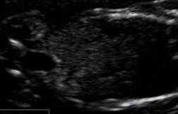
As pregnancy progresses, the bowel becomes more heterogenous, centrally located and well defined with echogenic walls and hypoechoic contents. Late in pregnancy discrete fluid filled loops of bowel can be seen in virtually all fetuses.
Individual segments of small bowel should not exceed approximately 7-10 mm in diameter and 15 mm in length.
Peristalsis within the fetal bowel can be seen as early as 16 weeks of gestation.
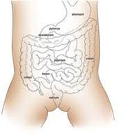
Normal large bowel
The large bowel is not clearly recognizable with prenatal sonography until the early third trimester. Thereafter it becomes increasingly easy to recognize.
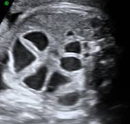
Jejunoileal atresia is commonly categorized as follows
- Type I – Membrane
- Type II – Blind ends joined by fibrous cord
- Type IIIa – Disconnected blind end
- Type IIIb – Apple-peel deformity
- Type IV – Multiple, string of sausages
|
|
JEJUNAL |
ILEAL |
|
|
|
|
|
|
|
|
|
|
|
|
|
|
|
|
|
|
|
|
|
|
|
|
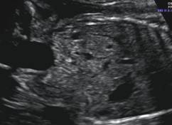
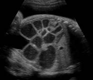
b. Meconium ileus
— Impaction of abnormally thick and sticky meconium in the distal ileum-functional obstruction
— Nearly all newborns have cystic fibrosis
— Dilated bowel segments containing echogenic meconium - usually seen after 26 weeks
— USG findings include
- Polyhydramnios
- Dilated bowel loops
- Meconium peritonitis
- Abnormal areas of increased echogenicity
— All the above findings are non specific and definitive diagnosis is difficult.
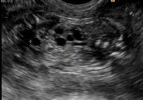
b. Hirschsprung disease
· Characterized by congenital absence of intramural myenteric nerve ganglia and sympathetic nerve plexus in a bowel segment.
· 1 in 10000 to 20000.
· Rarely presents prenatally, but when it does it is usually due to total colonic agangliosis.
· Sonographic features are non specific and rare. May be seen as dilated bowel segments and mild polyhydramnios
Bowel loops twist around the mesenteric artery or
ICOGU
CUSP XIV
What is the minimum duration for training in OB/GYN ultrasound?
View Results